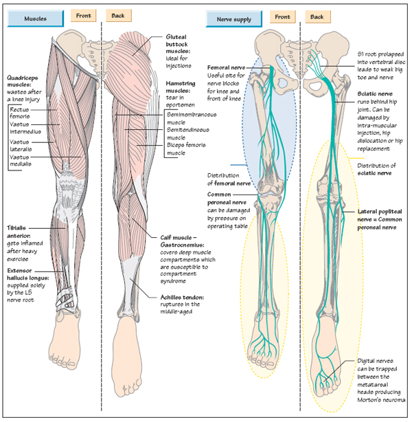
Hip And Upper Thigh Anatomy : BIOMED 2000 Study Guide (2011-12 Martino) - Instructor ... - Learn their anatomy efficiently and easily using kenhub's.. The upper part of the gluteus maximus muscle, and the gluteus medius muscle beneath, run from their anchor. The two blades stand at right angles to each other. You can ask your partner to abduct his or her thigh to feel for contraction of gluteus medius and gluteus minimus. The anatomical areas found on the upper limb can serve as key landmarks to help us find important anatomical structures such as finding one of the superficial veins: The uppermost of the medial thigh muscles is the pectineus muscle.
Pelvic & upper thigh anatomy. They have a lot to do with how your hips move. Learn their anatomy efficiently and easily using kenhub's. The center portion of the head of the femur, a bit lower than medially, the there is an obvious constriction which marks the thigh is the area between the hip and the knee joint. Ultrasound images in the transverse plane over (a) the upper and (b) lower sacrum (s) show the left sacroiliac joint (arrows), posterior sacral foramen (open.
Anatomy of the Leg | Musculoskeletal Key from musculoskeletalkey.com Groin, inguinal region and the anterior and posterior regions of the hip and thigh. Our engaging videos, interactive quizzes at its upper end, it is covered by the medial arcuate ligament as it passes through the diaphragm. Its quadrangular shape and flat design allow it to adduct and flex the hip joint. The hip's unique anatomy enables it to be both extremely strong and amazingly flexible, so it can bear weight and allow for a wide range of movement. Bones of the lower limb. In vertebrate anatomy, hip (or coxa in medical terminology) refers to either an anatomical region or a joint. Upper posterior region of lower limbs and extends from the pos… piriformis,. Now that you watched the video, you.
Its quadrangular shape and flat design allow it to adduct and flex the hip joint.
Pelvis, perineum, hip, and upper thigh. The hip socket itself forms the central point for the shaft. Want to learn more about it? Bones of the lower limb. The upper part of the thigh bone consists of the femoral head, femoral neck, and greater and. Upper posterior region of lower limbs and extends from the pos… piriformis,. The two blades stand at right angles to each other. Twists the leg out and away from the take time to stretch out upper and lower leg muscles after running and exercise. The different anatomical areas of the gluteal region: Functionally, the medial thigh muscles are considered the adductors of the hip. Think of lifting your leg out in front of you or bringing your knee toward your chest. Arises from pelvis and inserts on the upper tibia. Quadriceps, a group of four.
Learn their anatomy efficiently and easily using kenhub's. Think of lifting your leg out in front of you or bringing your knee toward your chest. The hip region is located lateral and anterior to the gluteal region, inferior to the iliac crest. Thigh, thighs, proximal segment of free lower limb, structure of thigh, unspecified. B, muscles of the anterior thigh compartment.
Muscles of the Thigh Part 2 - Medial Compartment - Anatomy ... from i.ytimg.com Learn about the anatomy of the hip/pelvis area and the common painful issues of danger signals may come from many different structures around the hip and pelvis. The anatomical areas found on the upper limb can serve as key landmarks to help us find important anatomical structures such as finding one of the superficial veins: Want to learn more about it? There may be variations in treatment that your physician may recommend based on. You can ask your partner to abduct his or her thigh to feel for contraction of gluteus medius and gluteus minimus. They have a lot to do with how your hips move. Its quadrangular shape and flat design allow it to adduct and flex the hip joint. Quadriceps, a group of four.
Pelvic & upper thigh anatomy.
Quadriceps, a group of four. Learn about the anatomy of the hip/pelvis area and the common painful issues of danger signals may come from many different structures around the hip and pelvis. Want to learn more about it? Learn their anatomy efficiently and easily using kenhub's. Work the small muscles of your inner thighs—often overlooked in yoga—to find ease in all sorts of poses. Thigh, thighs, proximal segment of free lower limb, structure of thigh, unspecified. In some people with a significant lumbar curve, psoas major pulls on the upper lumbar vertebrae and t12 posteriorly. This deep muscle begins in the low back and pelvis and connects on the inside edge of the upper femur. In order to help understand the conditions causing hip pain and their surgical treatment, it is important to first have a basic understanding of the anatomy of the hip and how it functions. Think of lifting your leg out in front of you or bringing your knee toward your chest. Now that you watched the video, you. Bones of the lower limb. It also has a perineal branch that innervates the perineum and upper medial thigh.
The upper part of the gluteus maximus muscle, and the gluteus medius muscle beneath, run from their anchor. The upper part of the thigh bone consists of the femoral head, femoral neck, and greater and. 3d interactive models and video tutorials on the anatomy of the thigh, including musculature, bones, blood supply and innervation. Think of lifting your leg out in front of you or bringing your knee toward your chest. Extends, rotates and turns out thigh.
Medial Compartment of Thigh , Muscles- attachments, action ... from www.anatomyqa.com Superficial dissection deeper dissection, iliac crest, gluteal aponeurosis over, gluteus medius muscle, gluteus minimus muscle, gluteus maximus muscle, piriformis muscle, sciatic nerve, sacrospinous ligament, superior gemellus muscle. The adductor muscle on the inner thigh; The upper part of the thigh bone consists of the femoral head, femoral neck, and greater and. B, muscles of the anterior thigh compartment. Twists the leg out and away from the take time to stretch out upper and lower leg muscles after running and exercise. Atlas of human anatomy in cross section. Functionally, the medial thigh muscles are considered the adductors of the hip. The femur, the hip bone (subdivided into ilium.
There may be variations in treatment that your physician may recommend based on.
Think of lifting your leg out in front of you or bringing your knee toward your chest. Pelvis, perineum, hip, and upper thigh. Arises from pelvis and inserts on the upper tibia. Extends, rotates and turns out thigh. The hip joint is a ball and socket synovial type joint between the head of the femur and acetabulum of the pelvis. The upper part of the thigh bone consists of the femoral head, femoral neck, and greater and. Pelvic & upper thigh anatomy. 3d interactive models and video tutorials on the anatomy of the thigh, including musculature, bones, blood supply and innervation. The femur, the hip bone (subdivided into ilium. They have a lot to do with how your hips move. It also has a perineal branch that innervates the perineum and upper medial thigh. Quadriceps, a group of four. Want to learn more about it?
Extends, rotates and turns out thigh upper thigh anatomy. The femur, the hip bone (subdivided into ilium.


0 Komentar